

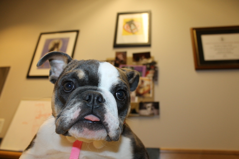
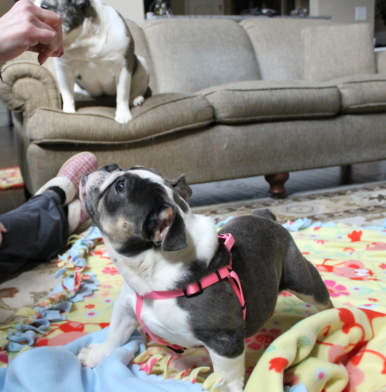
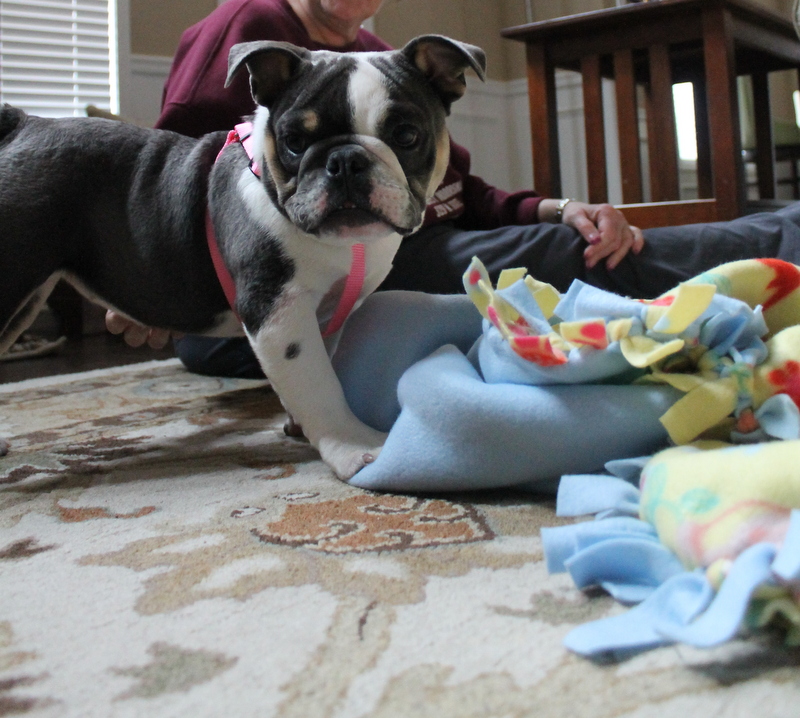
3/20/14: Please welcome Reagan! She came to us from a breeder by the way of our friend Melissa. Reagan was born with a heart murmur so of course she is no use to the breeder. But thank goodness he did not breed her or sell her to someone else that could breed her.
She had a visit with the cardiologist on 3/25. He confirmed she has two heart defects; a ventricular septal defect and pelmonic stenos but she is ok! (Notes from Dr. Pouge on both defects are below). At this time he does not feel she needs any medication and that these defects should not shorten her life span. She will need to see him again for another echocardiogram at 1 year of age as the pulmonic stenosis severity may get worse as she grows and treatment may be recommended at that time.
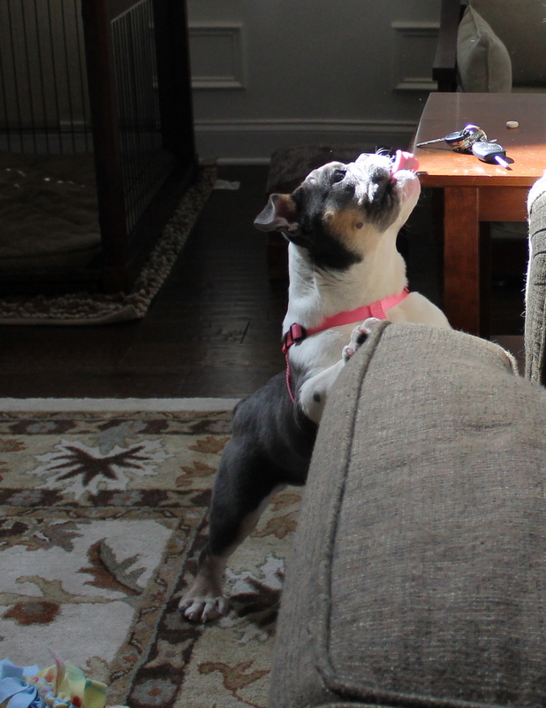
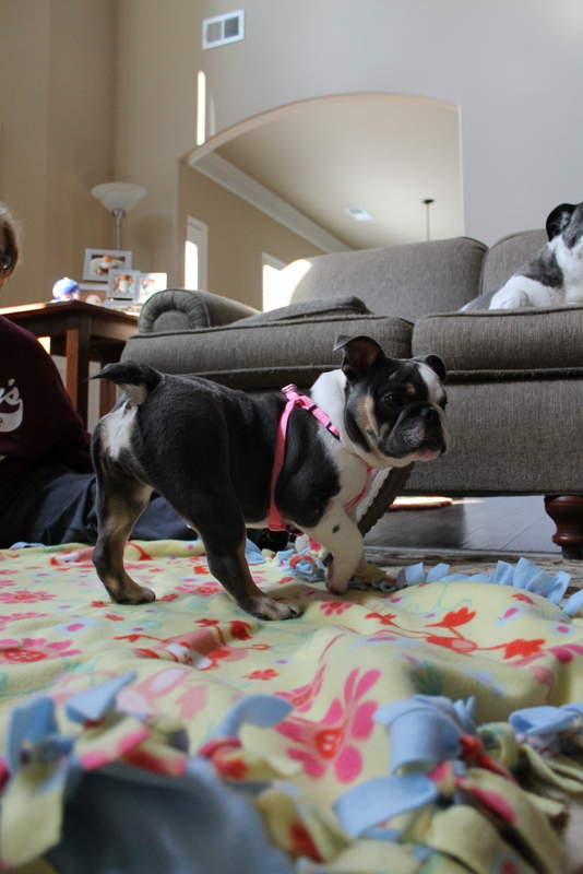
She is also a little weak in the legs still. Dr. Rush believes this is from spending the first four months of her life in a crate. We have noticed improvement in the short time she has been with us as she has been able to move around and be more active.
Dr. Rush spayed her and shortened her palate on 3/28 and she moved into her foster home on 4/3. She is up to date on her vaccinations but is being treated for mange. She will have a recheck in two weeks and if the mange has cleared up she will be available for adoption.
If her foster parents do not adopt her at the time then we will be accepting applications for her. Please note that all applicants must be local, be active with the rescue and commit to the echocardiogram with Dr. Pouge in eight months. Any and all costs associated with that procedure or any other procedures and/or medication she may need at the time will be the financial responsibility of her adopter not GEBR.
Reagan was born on 11/17/13. She is good with other dogs. We have not seen her around cats or children.
Update 5/14/14: Reagan is mange free and adopted! Her foster family fell in love with her and made it official. Congrats Reagan, Jen, Garett and Ginger, GEBR alum.
GUARDIAN ANGELS:
B & D Howell


Ventricular septal defect (VSD):
This defect is due to abnormal development of the ventricular septum which separates the left and right ventricles from each other. With a perimembranous defect (what Regan is affected with), there is a hole at the top of the septum, right under the aortic valve. The hole is very small and only a mild amount of blood is shunting from the left ventricle into the right ventricle and lungs. Because the hole is small and will not get any larger, I do not expect this defect to significantly affect her lifespan, though she will always have a murmur.
Pulmonic stenosis:
This condition is due to the abnormal development of one of the valves of his heart, the pulmonary valve. The pulmonary valve separates the right side of the heart (specifically, the right ventricle) from the large vessels of the lungs (main pulmonary artery) and is responsible for preventing the back-flow of blood between these two structures. With pulmonic stenosis, valvular malformation causes the opening between the right ventricle and the main pulmonary artery to be narrowed, which in turn obstructs the outflow of blood into the lungs. As a consequence, the right ventricle is forced to pump harder to force the same volume of blood through a narrowed orifice. This leads to thickening of the walls of the right ventricle (pressure hypertrophy). As blood rushes past the narrowed orifice and strikes the walls of the pulmonary artery past it, an audible murmur is created (as your veterinarian heard). In some animals with pulmonic stenosis, the abnormal conformation of the pulmonic valve causes it to be leaky, as well; this is known as valvular insufficiency.
The consequences of pulmonic stenosis are related to the severity of bloodflow obstruction and the severity of valvular insufficiency, if present. Currently the bloodflow obstruction is mild and as long as it stays within the mild/moderate range, it may not affect her length/quality of life and requires no specific procedures to open the valve more. If it becomes more severe as she grows, changes to the right side of the heart can lead to clinical signs (seen in approximately 35% of severely affected animals) such as fatigue, fainting, or fluid build-up in the abdomen and chest. These signs are usually seen in older animals with markedly abnormal pulmonary valves. Although exact criteria for the establishment of prognosis in dogs with pulmonic stenosis have not been created in animals, it is generally observed that dogs with mild pulmonic stenosis (which Regan has) are at low risk of clinical signs or problems throughout their life.
As we have discussed, English bulldogs commonly have an abnormality of their coronary vessels (those which run over the surface of the heart itself and supply oxygen to the heart muscle), know as an R2A anomaly. This abnormality is found in a large percentage of bulldogs (thought to be greater than 50%), and is important in that it complicates catheter procedures used to correct pulmonic stenosis. We have evaluated for the presence of this abnormality in Regan and she may have an abnormal coronary artery. However, an angiogram (performed during herprodecure) would be necessary to confirm this finding.
click photos to enlarge