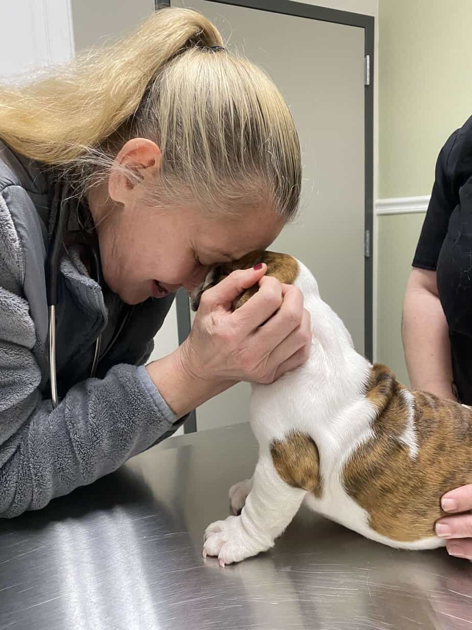
1/14/25 – Please welcome Francisco! He is a 9 week old puppy that was turned over to us by a breeder because he has a heart murmur. He will be seeing the cardiologist soon for a consult and surgery plan.
1/16/25 –
DIAGNOSTIC TESTING Echocardiogram:
1. Restrictive, perimembranous, left to right shunting ventricular septal defect: Characterized by a 2.9mm,
perimembranous defect. VSD Vmax 4.94m/s
2. Pulmonary valve dysplasia with mild pulmonary insufficiency: Intermittent elevated PVmax (1.8m/s).
3. Equivocal pulmonary artery enlargement: DDx. variant of normal vs. secondary to overcirculation.
ASSESSMENT:
Francisco, a 2-month-old male intact English Bulldog, was presented to the UGA Cardiology Service on 1/16/25 as a referral from his primary care veterinarian following auscultation of a grade V/VI heart murmur. On physical examination today, a grade V/VI basilar systolic murmur (R>L) was heard, and an echocardiogram was recommended to further evaluate for a structural abnormality in Francisco’s heart.
On echocardiogram, we confirmed the presence of a left to right shunting ventricular septal defect (VSD). A ventricular septal defect (VSD) is a congenital abnormality in which the interventricular septum (muscular wall between the left and right ventricles of the heart) is incompletely formed, resulting in inappropriate movement of blood from the left (oxygenated) chamber into the right (de-oxygenated chamber). Fortunately, the majority of VSDs are small and do not cause significant cardiac compromise, as is the case with Francisco. Clinical findings depend on the severity (size) of the defect in the muscular wall and the direction of blood flow, but a small, restrictive defect usually causes minimal or no clinical signs. Further, most minor cases of VSD do not require any treatment and patients go on to have a relatively normal lifestyle and life expectancy. VSDs typically stay the same size or may spontaneously close in some cases. However, if Francisco’s VSD were to become larger he may begin to exhibit secondary changes to the heart (enlargement of the left side of the heart) due to increased workload. Possible sequelae of large VSDs include pulmonary hypertension (elevated pressure in the lungs) or congestive heart failure (backup of fluid into the lungs). In extreme cases, the pressure in the lungs could become so great that the flow of blood through his VSD could reverse, and deoxygenated blood could be circulated throughout the body. Should his condition worsen, physical occlusion (closure) of the defect would be indicated. This could be achieved via intravenous placement of an occluder device or an open heart procedure. Francisco is not currently exhibiting any signs of cardiac compromise, and we expect he will continue to do well.
In addition to the VSD, we also noted that Francisco has a very mild malformation of his pulmonary valve, which we refer to as “pulmonary valve dysplasia”. Pulmonary valve dysplasia is a congenital heart defect of the pulmonic valve, which is located between the right ventricle and the pulmonary artery (the great vessel that takes blood to the lungs). In Francisco, the leaflets of this valve are thickened and partially fused together.
There is also evidence of mild pulmonic insufficiency due to dysplasia (malformation) of his pulmonic valve. Therefore, during diastole (ventricular filling period), there is backflow of blood into the right ventricle. These changes in Francisco are mild at this time, and should not result in any significant cardiac changes. We will continue to monitor this abnormality in subsequent echocardiograms.
AT-HOME INSTRUCTIONS:
ANESTHESIA:
There are no major cardiac structural changes at this time as a result of Francisco’s ventricular septal defect. However, given that Francisco has a shunt present, general anesthesia carries the risk of altering pressures within the heart, which could result in the shunting of deoxygenated blood into the systemic circulation. As a result, we recommend avoiding the use of ketamine and alpha 2 agonists (i.e. dexdomitor) in the anesthetic protocol, which could significantly alter blood pressure. Monitoring with ECG, blood pressure, and SpO2 is recommended. Further anesthetic recommendations can be obtained through the UGA Anesthesiology service.
GENERAL MONITORING
Please monitor Francisco for development of symptoms of heart disease or failure. These may include coughing, rapid or labored breathing, abdominal distension, lethargy, exercise intolerance, or episodes of collapse (fainting or sudden weakness). If any of these symptoms are observed, please call us immediately to discuss an appropriate plan.
SLEEPING RESPIRATORY RATE
Please monitor Francisco’s sleeping respiratory rate as a possible indicator of congestive heart failure. As previously discussed, you can obtain this rate by counting the number of times Francisco takes a breath (either in or out, whichever you find easier) in 15 seconds and multiplying that number by 4, or by using the Cardalis Health & Fitness iPhone app. If you obtain a number higher than 30 (best to repeat a few times to make sure it is “real”), or if you notice an upward trend compared to Francisco’s usual rate, please call us to discuss whether a medication change might be necessary.
EXERCISE
As a general rule, Francisco can be allowed to determine his own activity level. Extreme levels of activity should be avoided, and Francisco should not be pushed to do more than what he wants to do, but normal amounts of activity and play are fine.
FOLLOW-UP:
in 4-6 months to recheck echocardiogram, then again in 6 months.
1/24/25 – Francisco had been adopted. Congrats Jameson, Roger and Cristi!
Bully Bag Sponsors: Carol Robertelli, Derek and Brenda Berube
Name That Bully Sponsor: Brendan and Jen Magee