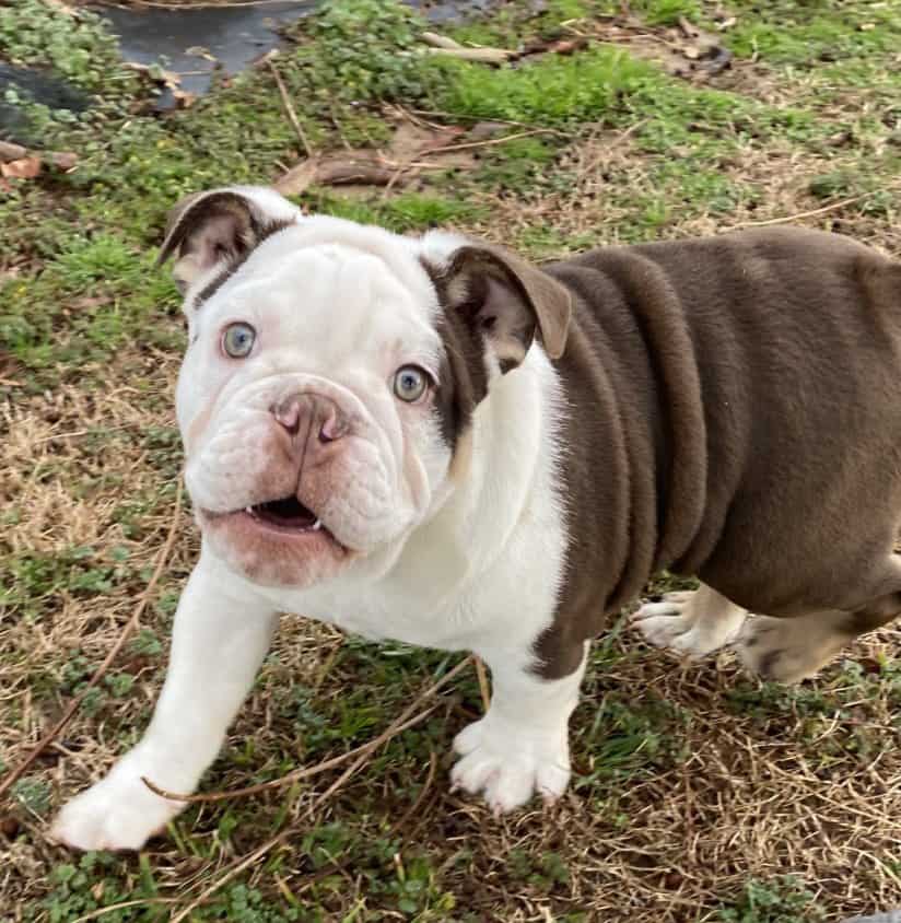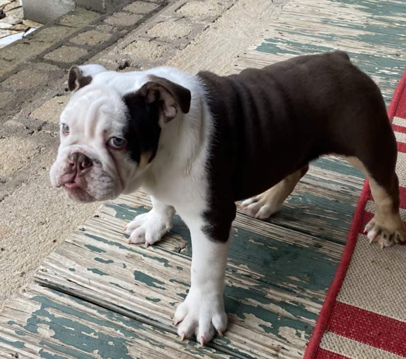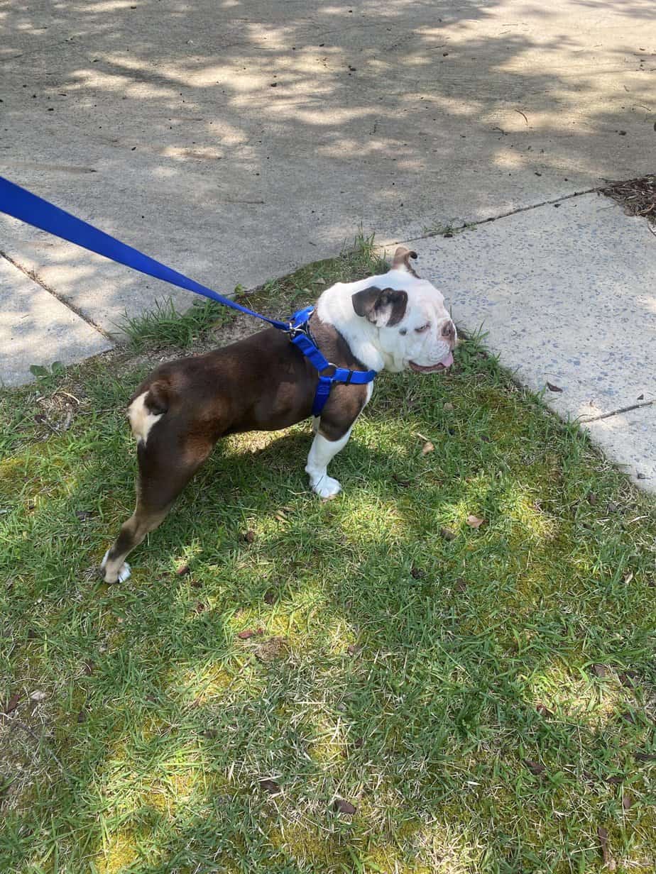

5/8/23 – Please welcome Cowboy! Cowboy was born on 11/22/22 with a heart defect and turned over to us by his breeder. He had an initial work up with the cardiologist in February and started showing signs of struggling recently. We started him on atenolol today and he will see the cardiologist at UGA in a few weeks for further testing. Atenolol is a drug is in a class of drugs called “beta-blockers.” We are using it primarily to decrease the strength of contraction, which may be beneficial for Cowboy’s type of heart disease. It will also have the effect of decreasing heart rate, which gives the heart more time to relax.
6/1/23 – Cowboy saw the cardiologist today. The echocardiogram shows he has moderate pulmonary valve stenosis, with a mean peak pressure gradient of 59 mmHg. There is secondary remodeling of the right heart, with moderate right ventricular concentric hypertrophy, mild to moderate right atrial dilation, and mild to moderate right ventricular eccentric hypertrophy. The pulmonary valve annulus measured approximately 11 mm. There is a small (4 mm), bidirectional (primarily left to right with a VSD PGmax of 17.5 mmHg) perimembranous ventricular septal defect.
ASSESSMENT:
Pulmonary valve stenosis is a congenital heart defect of the pulmonary valve, which is located between the right ventricle and the pulmonary artery (great vessel that takes blood to the lungs). In dogs with pulmonary valve stenosis, the leaflets of this valve are mildly thickened and/or partially fused together. In normal dogs, blood flows from the right ventricle through the pulmonary valve, to the pulmonary artery, and finally to the lungs to get oxygen. In Cowboy’s case, however, the pulmonary valve leaflets are fused together, and they do not open all the way when his heart pumps blood forward. His outflow is also mildly narrowed (annular hypoplasia). Because of this, the right side of the heart has to work harder to pump blood out of the heart and into the lungs due to the abnormal valve. Because of this increased work load placed on the right heart, the heart muscle has remodeled (become thickened) as well.
Fortunately, Cowboy’s pressure gradient is in the moderate category, and at this time he only has mild to moderate changes to his right ventricle. At this time we do not believe he needs to have a balloon valvuloplasty to alleviate his pulmonary valve stenosis. Instead we will continue him on atenolol at this time.
Cowboy also had a ventricular septal defect today on echocardiogram. A ventricular septal defect is a congenital heart defect that allows abnormal shunting of blood between the two lower chambers of the heart, the left and right ventricles. Usually, these chambers receive blood from the body (the right atrium/ventricle) or the lungs (the left atrium/ventricle), and are separated by a large wall, called the inter-ventricular septum. This wall is important, because it prevents the mixing of poorly oxygenated blood from the tissues of the body with the well-oxygenated blood returning to the heart from the lungs.
Often patients with VSDs do not show symptoms; however, if the defect is large, the heart can become overwhelmed by the increased workload and eventually develop congestive heart failure. In a typical VSD, well-oxygenated blood moves from the left side of the heart to the right side of the heart. However, when the heart is overloaded, the shunt can switch to a right to left shunt. This means that poorly oxygenated blood is now being pushed into systemic circulation, meaning the rest of the body is not receiving enough oxygen. Clinical signs seen are collapse, inability to sustain normal activity levels, difficulty breathing, cyanosis (blue- ish discoloration of the skin). Small ventricular septal defects generally do not require treatment. Fortunately, this defect appears small for Cowboy at this time, but the flow of blood is bidirectional (although primarily left to right). This could result in de-oxygenated blood going into circulation. With this, it will be important to monitor his red blood cell levels (PCV), which was normal today. As these dogs push more de-oxygenated blood to their body, it signals to their body to produce more red blood cells resulting in an excess amount. If his blood cells get too high, intervention may be required. This would essentially be removing some of his blood and replacing it with fluid to essentially dilute his blood cells. For larger defects, they are often treated with vasodilators to lower the blood pressure and decrease the amount of shunting, as surgery to close the defect is not widely available.
At this point we are comfortable continuing Cowboy on atenolol, in hopes that it will reduce the workload on his heart. If at future echocardiograms Cowboy has progressed into the severe category for his pulmonary valve stenosis, or he has more severe changes to his right ventricle, we may recommend a balloon valvuloplasty at that time. He will see the cardiologist again in six months.
12/4/23 – Cowboy saw the cardiologist for his follow up today and it was determined that we should move forward with the balloon valvuloplasty.
12/6/23 – Cowboy had his surgery today and all went well. He will be on strict rest while he recovers over the next two weeks but we suspect he will make a full recovery and live a long healthy life. He will see the cardiologist in four months for a recheck.
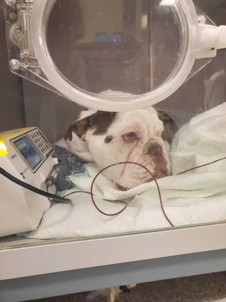
3/23/24 – Cowboy was lethargic today so we got him to the emergency clinic for observation. He is having trouble breathing on his own so we have him in the oxygen chamber. We will move forward with bloodwork and x-rays when he is stable.
3/24/24 – We called the cardiologist in to do further testing. We suspect his VSD has gotten larger and it is causing issues. At this time his red blood cell count is extremely high and we suspect his body is not oxygenating his blood properly. Unfortunately we had to make the tough decision to let him go.
March 24, 2024 – Cowboy crossed over the Rainbow Bridge today. Please keep his foster family in your thoughts. Rest In Peace Cowboy