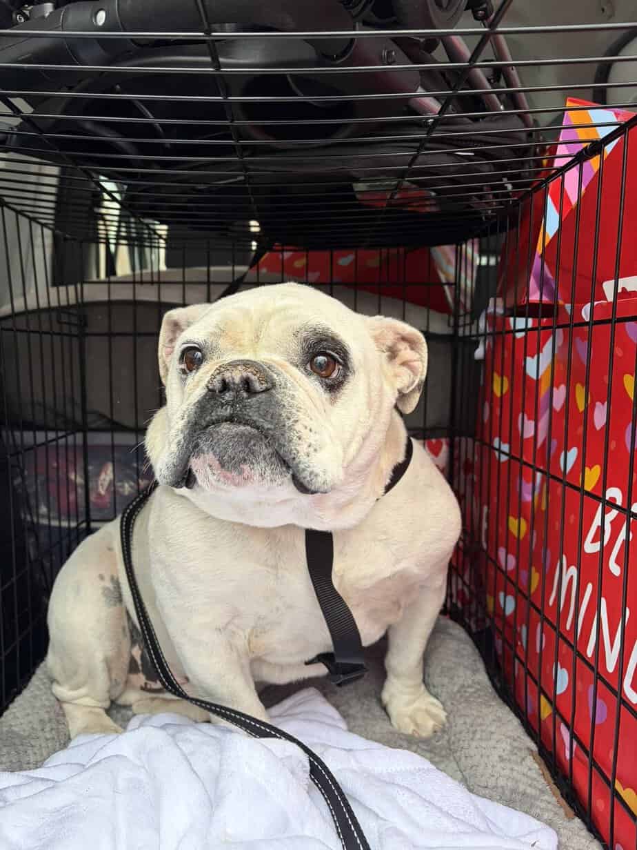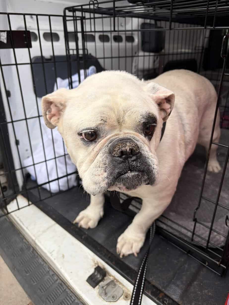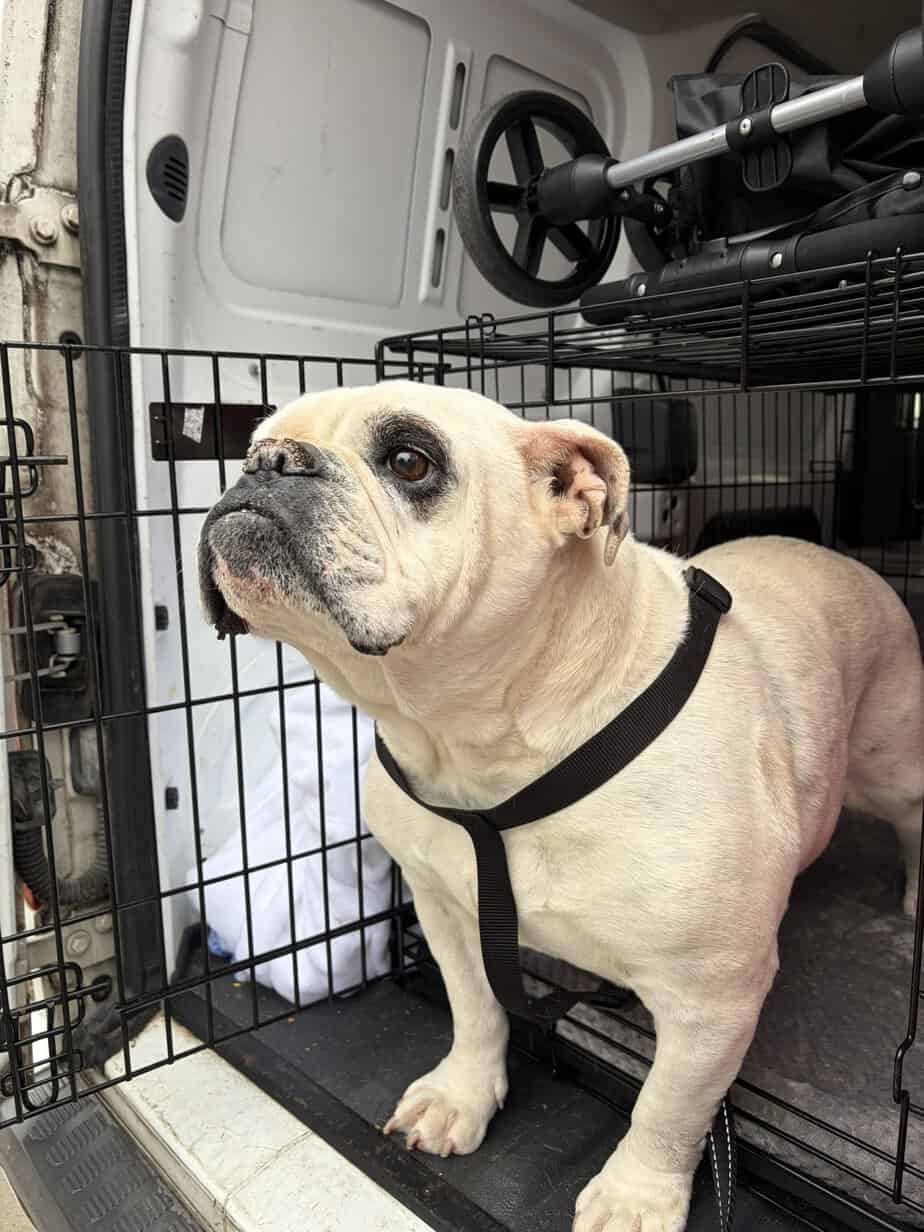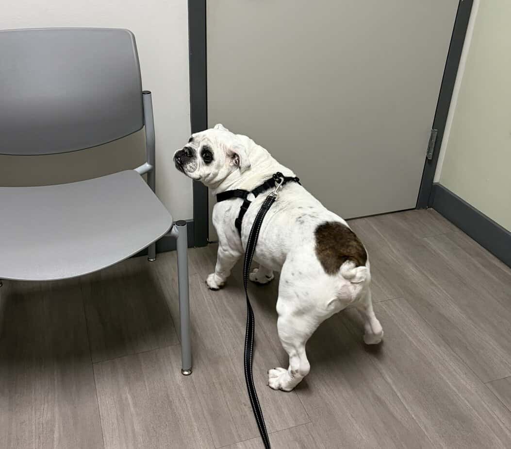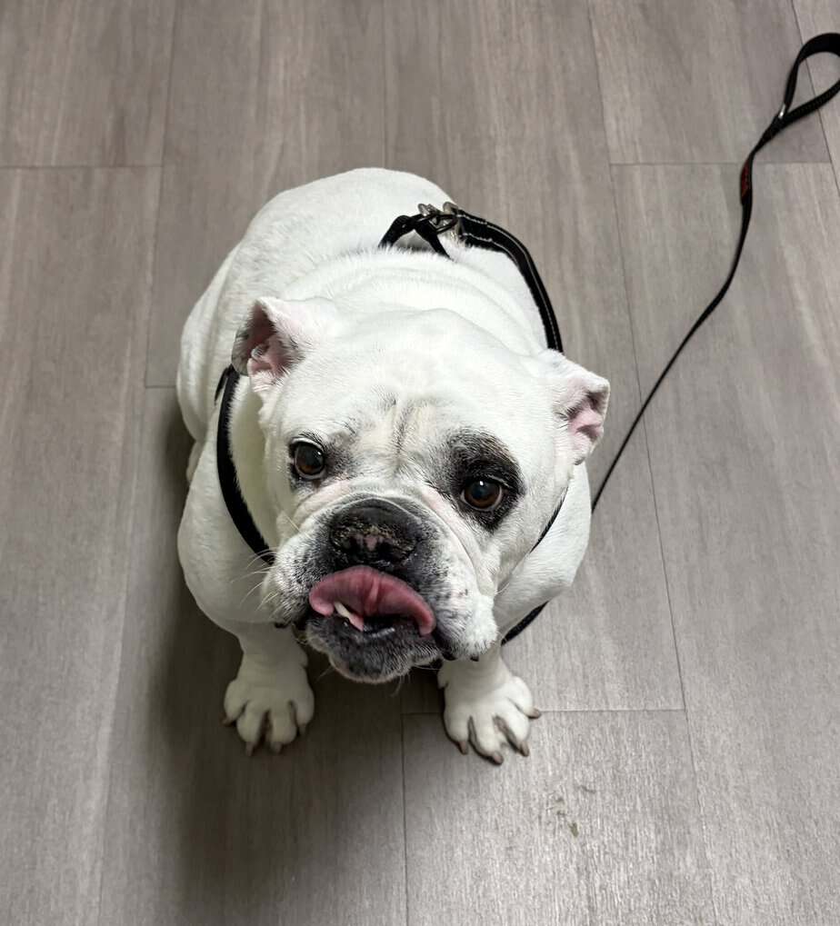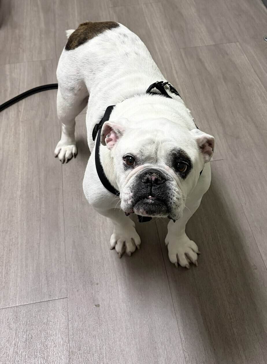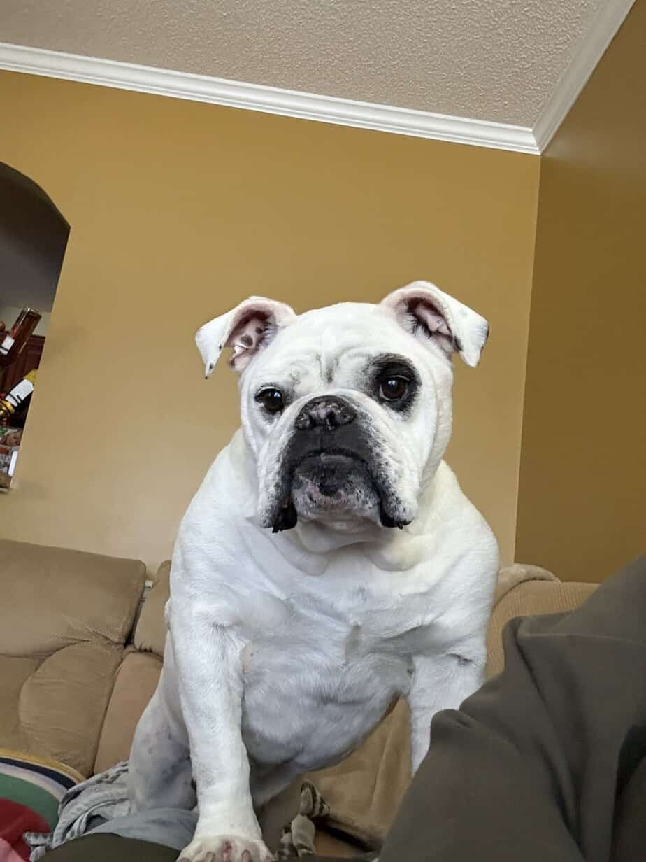
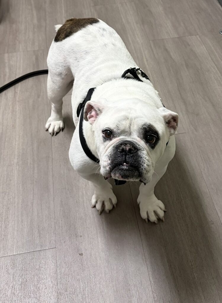
1/21/26 – Please welcome Capone! Capone’s owners were facing some health issues, so they asked us to find Capone a new home. He is already neutered and seems to be well cared for. He does need to drop some pounds so that he will be at a healthy weight.
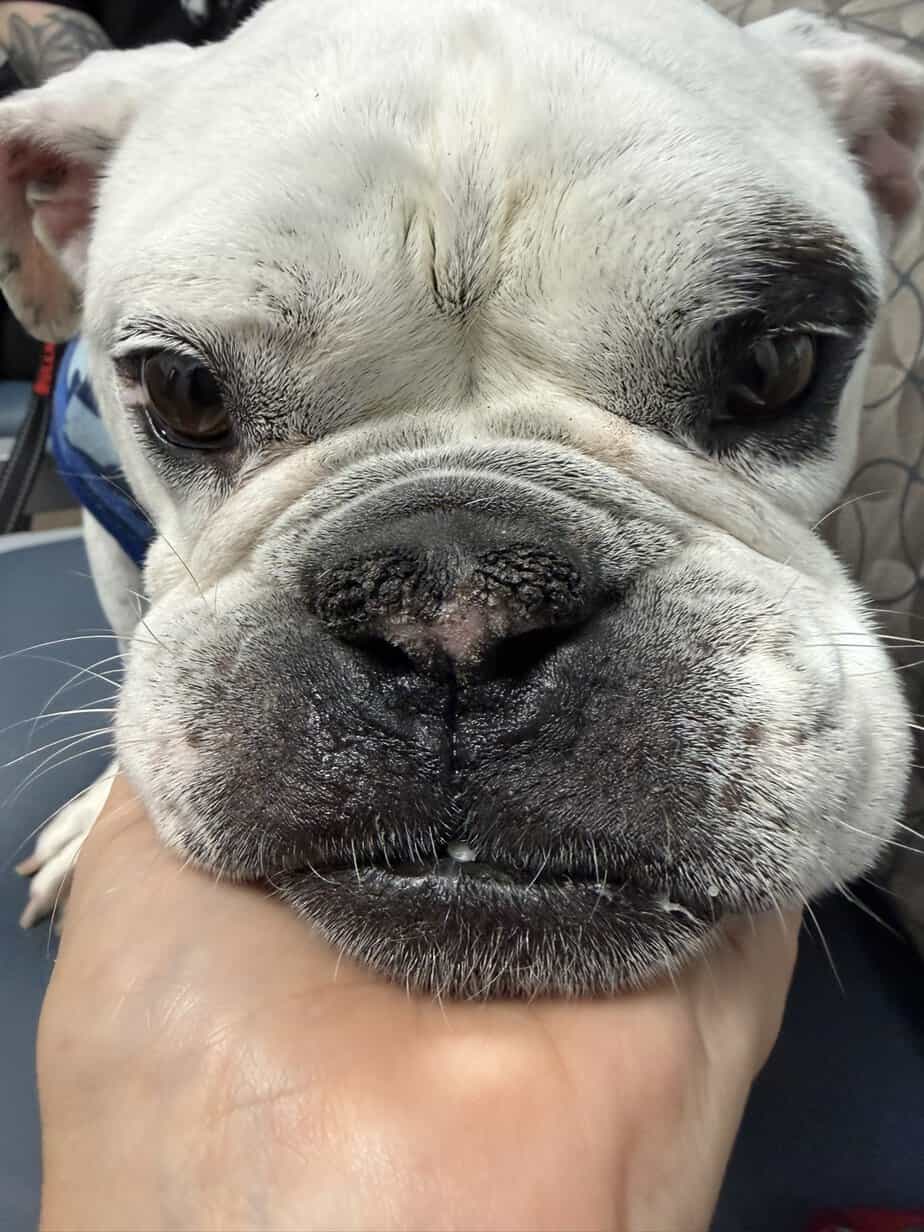
2/17/26 – Capone developed a mass that has ulcerated and needs to be removed and send off for biopsy.
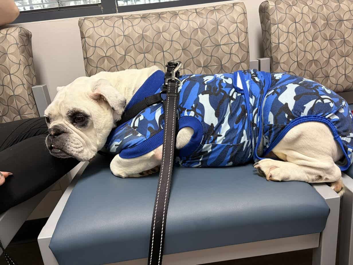
2/19/26 – Capone had surgery today and all went well. He had two masses removed, his palate shortened, his nares widened and his teeth cleaned.
3/3/26 – The biopsy report showed that one of the masses was a Squamous cell carcinoma. We will consult with the oncologist and do further testing to make sure nothing has spread.
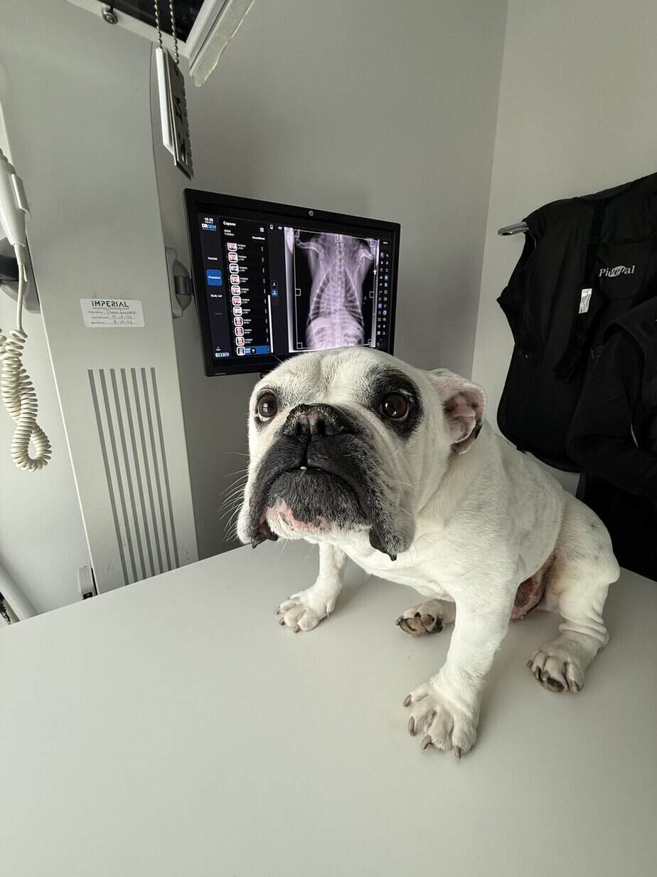
3/10/26 – Capone saw the oncologist today.
ASSESSMENT & PLAN
Squamous cell carcinoma (SCC) can be seen in the skin, and are usually locally aggressive, meaning they can grow to large sizes, can become ulcerated and painful, and can even invade into muscle and bone. Metastasis (cancer spread) is luckily uncommon. Prolonged outcomes of over two years are reported for dogs with inguinal (groin) SCC, but pets with higher grade tumors or vascular invasion may have poorer prognoses. Benefit from chemotherapy is not well defined. We know that Capone has evidence of vascular invasion on his pathology report, meaning cancer cells were seen by the pathologist in the blood vessels surrounding
his tumor. Abdominal ultrasound was performed today to rule out cancer spread (metastasis). We did note a splenic mass on ultrasound today, and samples were taken and submitted for cytology (pathology evaluation) to ensure this is not representative of cancer. Otherwise, no evidence of metastasis was noted on abdominal ultrasound today, nor on chest x-rays performed by your primary care veterinarian. I do not recommend chemotherapy for Capone, as SCC does not respond well to chemotherapy, even with the finding of vascular invasion. Pending the splenic cytology results, I would consider recheck abdominal ultrasound and physical exam
every 6-12 months since vascular invasion was noted.
3/15/26 – We received the biopsy on the fluid tacken from the mass in the spleen. The good news is that it is just a benign splenic mass, no concerns and no need to remove the spleen based on these results. We can monitor the size of the mass, but it is not uncommon to see benign splenic masses in older dogs. The spleen is a secondary site of red and white blood cell production outside of the bone marrow, and sometimes it can cause these spots to develop. Right now, the recommendation to recheck exam and ultrasound in six months and he will likely need exams and ultrasounds every 6-12 months to monitor his skin and spleen.
PATHOLOGY
CYTOLOGY WITH MICROSCOPIC DESCRIPTION (1 SITE/LESION)—STANDARD
Cytology Source: a SPLEEN
Clinical History: History of an cutaneous inguinal squamous cell carcinoma removed from the left inguinal region on 2/19/26. Vascular invasion noted on histopath. Incidental splenic mass noted on ultrasound for staging. No inguinal or iliac lymphadenopathy.
Pathology Report
INTERPRETATION:-Negative search for squamous cell carcinoma metastasis-Extramedullary hematopoiesis (normal)-Possible lymphoid hyperplasia (immune stimulation)
COMMENTS: No evidence of a squamous cell carcinoma metastasis in the area sampled. Extramedullary hematopoiesis is often seen in splenic tissue of normal individuals and is poorly specific but can be more prominent with anemia. Lymphoid cells predominate in some areas of the smears, which may simply be due to aspiration of a hyperplastic lymphoid follicle. This is usually associated with immune stimulation but also remains non-specific. When there is this combination of cytological findings in a splenic mass, fibrohistiocytic nodules and mesenchymal cell tumors can never completely be ruled out based on cytology as they can exfoliate their mesenchymal tissue poorly. Monitoring of this mass with repeat ultrasounds or a biopsy may eventually be needed for a more definitive diagnosis and prognosis.
CYTOPATHOLOGIC DESCRIPTION: The slides are of normal cellularity for splenic tissue. Few myeloid and erythroid precursors are seen and both series appear to show proportionate maturation sequences. They are intertwined with few clusters of small stromal cells. Many lymphoid cells also noted. Approximately 80-90% of the lymphoid cells are small mature lymphocytes. Less than 15% prolymphocytes and lymphoblasts are found. Rare plasma cells noted. Small amounts of hemosiderin are intertwined with the few clusters of splenic stroma found throughout the smears. Rare hemosiderin laden macrophages also noted. Rare heavily granulated mast cells are noted. The background consists of peripheral blood, tissue
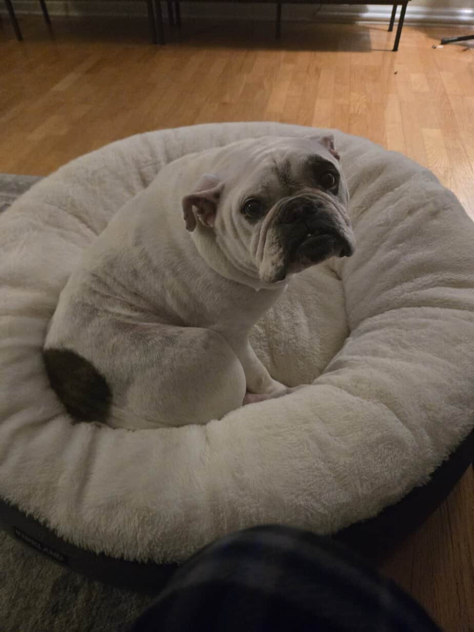
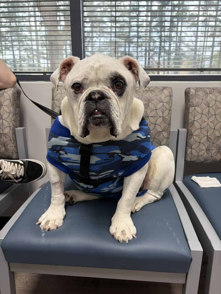
3/27/26 – Capone has been cleared for adoption. We understand he will need additional care for the rest of his life to monitor his skin and spleen but we know whoever adopts him wont mind one bit. He is a sweet fella and doing well with the recent changes in his life!
approximate age: 7 – DOB 5/1/2018
approximate weight: 63, down from 67 when he came into rescue
likes dogs: we are told from his previous owner that he likes them, he is not currently living with any in his foster home, he saw a few at the vet clinic and did not mind them but he saw a few at a public event and he seemed to be upset by them
likes cats: we are told from his previous owner that he likes them
likes kids: we are told from his previous owner that he likes them
food: Taste of the Wild, Pacific Stream
lifelong medications: none
likes: cuddling, playing and a little wrestling with his people
dislikes: water sprinklers in the yard
ideal home: we think he would fit well in just about any home but would love to have people close by him as often as possible. He loves human contact, and is a very sweet animal
Bully Bag Sponsors: Carol Robertelli, Stefanie Slay